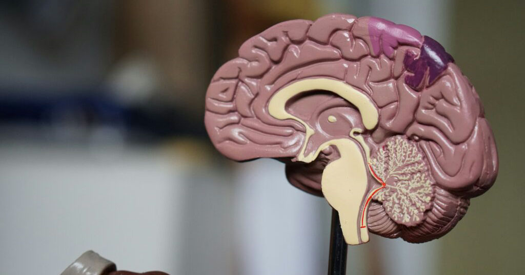
Brain Imaging & Neuro Epidemiology
The BraINE group studies brain health by exploring the factors that increase or decrease the risk of brain diseases. They also develop methods to predict and detect these diseases at an early stage.
ACTIVITIES
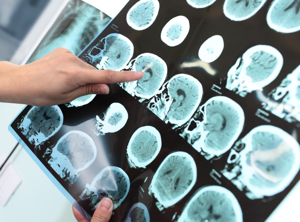
Research focuses on both healthy individuals and patients, as well as animal models to better understand different diseases. The team primarily uses advanced brain imaging technologies (fMRI, MRI, PET), but they also study how memory and cognitive abilities are influenced by various factors, beyond just the brain’s structure.
With a strong focus on improving patient care, the BraINE group develops advanced methods for analysing medical images, using radiomics (which extracts detailed information from images) and deep learning (artificial intelligence techniques that help detect complex patterns in the images).
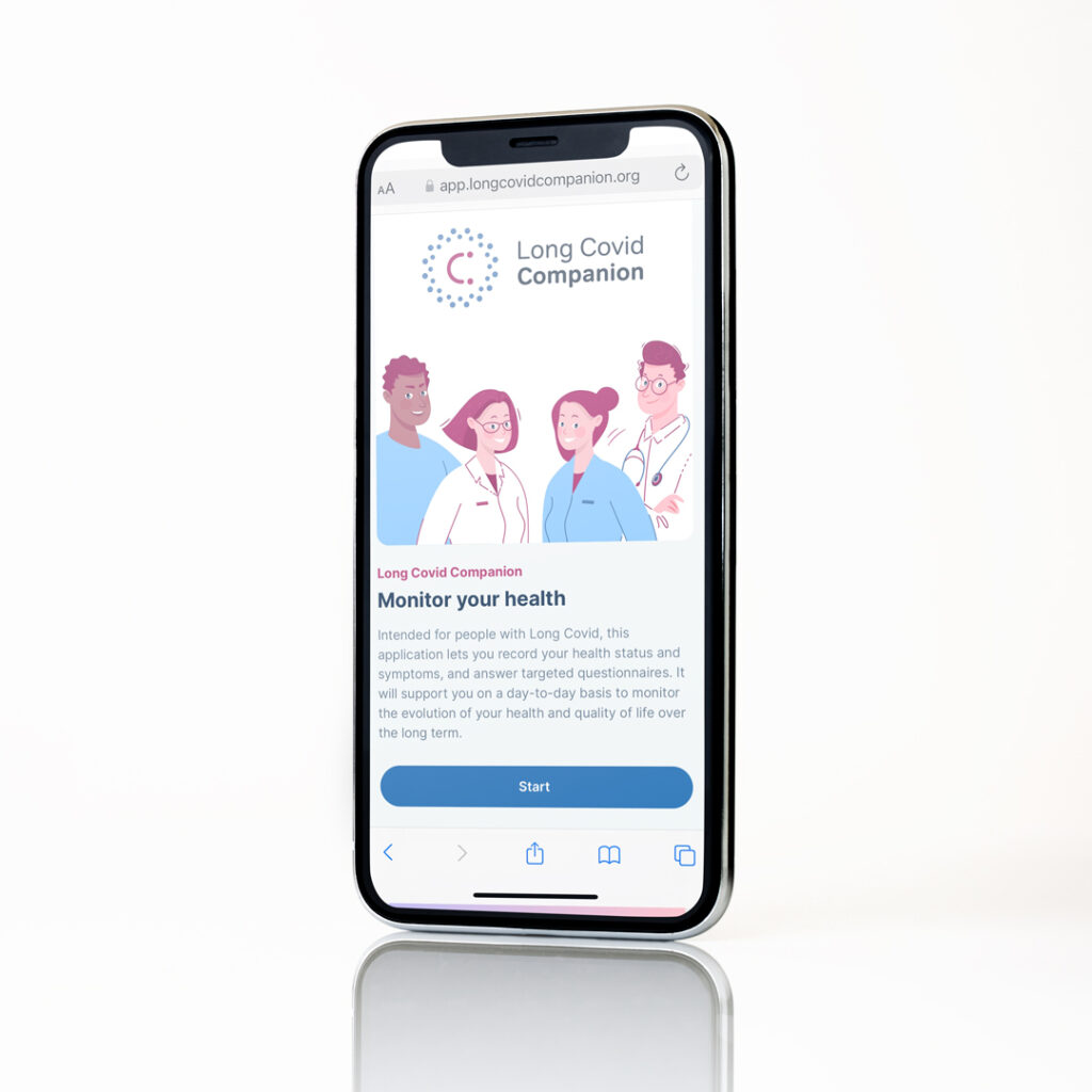
Current projects focus on brain diseases that worsen over time, such as brain tumours, Alzheimer’s and Parkinson’s, as well as the long-term effects of COVID-19 on the brain.
The team is affiliated with both the Department of Cancer Research and the Department of Precision Health at the LIH.


Partners & Funding


Projects & clinical trials
Featured team members
-
 Selma
Selma
Boudissa Research Engineer -
 Salah
Salah
Ghamizi Postdoctoral Fellow -
 Georgia
Kanli Research Engineer -
 Olivier
Keunen Group Leader -
 Elena
Elena
Lacomba Arnau Postdoctoral Fellow -
 Valérie
Palissot Scientist -
 Magali
Perquin Scientist
Scientific publications
-
Quantitative pre-clinical imaging of hypoxia and vascularity using MRI and PET – 01/01/2024
-
Simultaneous Image Quality Improvement and Artefacts Correction in Accelerated MRI – 23/10/2024
-
Multi-parametric MRI to FMISO PET Synthesis for Hypoxia Prediction in Brain Tumors – 09/10/2024
-
Addressing Artefacts in Anatomical MR Images – 01/05/2024
-
Early-life influenza A (H1N1) infection independently programs brain connectivity, HPA AXIS and tissue-specific gene expression profiles – 11/03/2024
-
Corrigendum to ‘Molecular Identity Changes of Tumor-Associated Macrophages and Microglia After Magnetic Resonance Imaging–Guided Focused Ultrasound–Induced Blood–Brain Barrier Opening in a Mouse Glioblastoma Model’ [Ultrasound in Med & Biol. 49 (2023) 1082-1090, (S0301562922006664), (10.1016/j.ultrasmedbio.2022.12.006)] – 01/01/2024
-
Predicting Hypoxia in Brain Tumors from Multiparametric MRI – 25/01/2024
-
Immune cell identity behind the Ktrans mapping of mouse glioblastoma – 21/06/2023
-
Molecular Identity Changes of Tumor-Associated Macrophages and Microglia After Magnetic Resonance Imaging–Guided Focused Ultrasound–Induced Blood–Brain Barrier Opening in a Mouse Glioblastoma Model – 01/01/2023
-
Author Correction – 26/01/2023
Job vacancies
-
2 Post-doctoral Researchers in Foundation Models for Neuro Imaging (MC/PDTR125/OK/TRSRD)
Department of Cancer Research – Research – Translational Radiomics (TRANSRAD)